Stony Brook University Biomedical Engineering

Biomedical Imaging Research Laboratory

BIRL · Stony Brook University

Research

Methods
Ultrasound elastography, functional ultrasound, photoacoustics and radiation-acoustic imaging, built on mechanics, inverse problems and physics-constrained learning, and carried from simulation through phantoms and animal models to patients.
Impact
Imaging biomarkers for cancer therapy response; functional ultrasound for mapping brain activity and following neurological disease; mechanical and microstructural markers in the ageing and HIV-affected brain; and verification of delivered dose in radiotherapy.

Focus areas

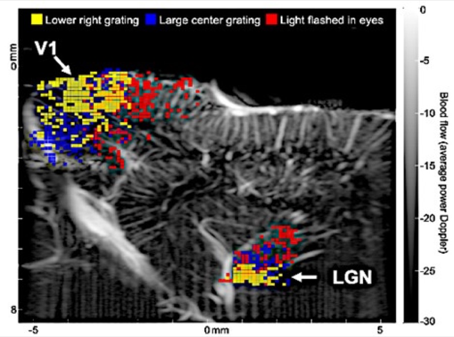
The figure shows fUS imaging of brain response to sensory stimuli. Gray map shows baseline image in sagittal plane of a ferret brain, including visual cortex (V1) and thalamus (LGN). Note the detailed vasculature. Following 30 seconds of patterned visual stimuli, increased signals were detected in V1 and LGN. Differently colored pixels show the highest activation correlated with different stimuli.

Functional Ultrasound

Functional magnetic resonance imaging (fMRI) is the current “gold standard” for human and primate brain imaging. However, employing fMRI in small animal research is challenging and very expensive. Additionally, fMRI’s low spatiotemporal resolution limits its effectiveness for small animal research. To deal with these issues, more researchers are now relying on optical imaging for basic neuroscience research in animals. Optical imaging has many attractive attributes, such as exceptional subcellular level spatial resolution and fast temporal resolution. However, the main challenge is that it cannot noninvasively visualize deep brain structures. Neuroimaging requires a different approach to tackle its main challenges, one that takes a fresh look at the critical issue – neither optical microscopy nor fMRI is ideal for live imaging of small animals’ brains. High frequency functional ultrasound (fUS) imaging is filling an essential gap in currently available functional brain imaging technology. fUS provides sufficient spatial and temporal resolution (~100 microns and tens of milliseconds) to image the activity of neurons while also enabling penetration into deep brain structures. Plane-wave imaging, the technology that underpins fUS, allows excellent separation of stationery and blood flow signal in small vessels from the surrounding tissues, which enables fUS to detect neural activity changes through neurovascular coupling. We are using fUS to study and understand the visual system.
Machine Learning in Medical Imaging

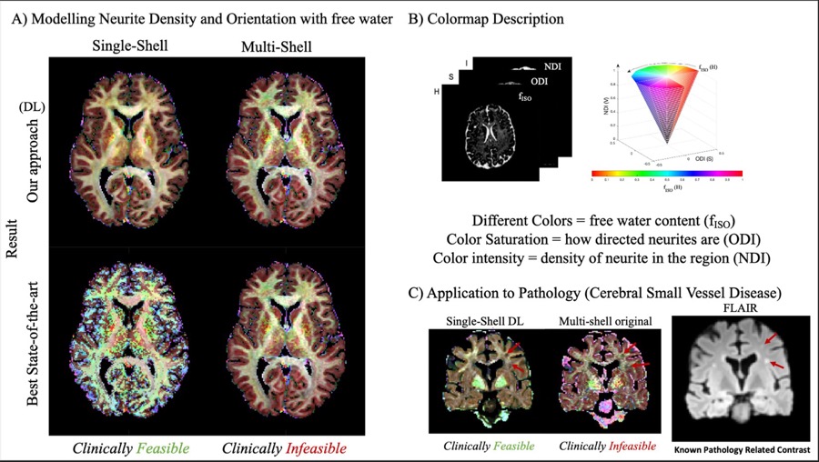
Noninvasive micro-structural investigations in clinical settings for any medical imaging modality enable the examination of unexplored pathological alterations. Artificial intelligence (AI) with data-driven strategies (i.e., machine learning/deep learning) can play a major role in this regard. Clinical datasets pose unique limitations and challenges as the data-driven techniques are often limited by inherent hallucination, ill-posedness, and lack of training samples. Thus it is important to optimize AI architectures specifically to clinical needs in order to maximize priors. Maximum likelihood estimator, Bayesian, and inherent geometrical organizational frameworks can be leveraged to develop additional priors to reconstruct objectively accurate micro-parameter maps of the brain using these data-driven strategies. Specific to diffusion magnetic resonance, one of our strategies shows a reconstruction of neurite density, dispersion, and free water brain map reconstructed with a clinical dataset with high objective accuracy.

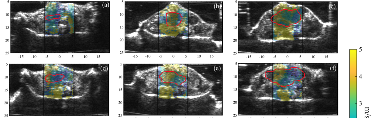
The figure shows shear wave speed overlaid on B-mode images of rectal tumors, with the tumor outlined in red.

Cancer Imaging

Response to cancer therapy is usually judged from tumor size, which can lag weeks behind the biology. We develop shear wave elastography and photoacoustic biomarkers that read the tumor microenvironment directly, measuring stiffness, attenuation and perfusion so that response to neoadjuvant therapy can be assessed early enough to act on. The work spans pancreatic and colorectal cancer in preclinical models.

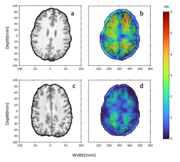
The figure shows anatomical images and the corresponding shear modulus maps for a 30-year-old and a 69-year-old participant.

Reverberant MR Elastography

Reverberant MR elastography measures the mechanical properties of the brain by exploiting the multi-directional wave field created by reflections within the skull, rather than requiring assumptions about wave propagation direction or boundary conditions. We use it to study how brain stiffness changes across adulthood, and to ask which microstructural changes underlie that decline by pairing elastography with diffusion measures of neurite density, orientation dispersion and free water. A related line relates brain stiffness to learning and memory deficits in people with HIV.

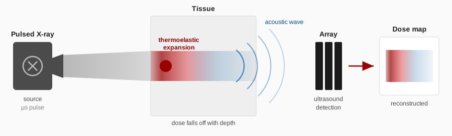
The figure shows the principle: a pulsed X-ray beam deposits energy in tissue, the resulting thermoelastic expansion launches an acoustic wave, and an ultrasound array detects it so the deposited dose can be reconstructed.

Radiation-Acoustic Imaging and Dosimetry

Radiotherapy delivers a dose that is carefully planned but rarely verified inside the patient. The laboratory is developing a prototype system for FLASH radiation-acoustic tomography (FRAT), which uses the acoustic signal generated as pulsed radiation deposits energy in tissue to map where that dose actually lands. Visualizing dose at depth matters most in FLASH radiotherapy, where very high dose rates and few fractions mean that small positioning errors carry large clinical consequences.

Related work continues in Elastographic Imaging · Harmonic Imaging · Photoacoustics · Ultrasound Beamforming.